488 Nm Laser Flow Cytometry

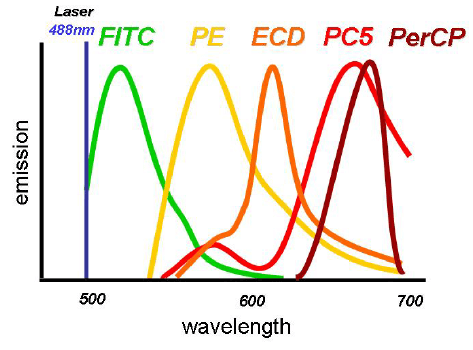
Flow Cytometry Facs Fluorochrome Selection Sino Biological

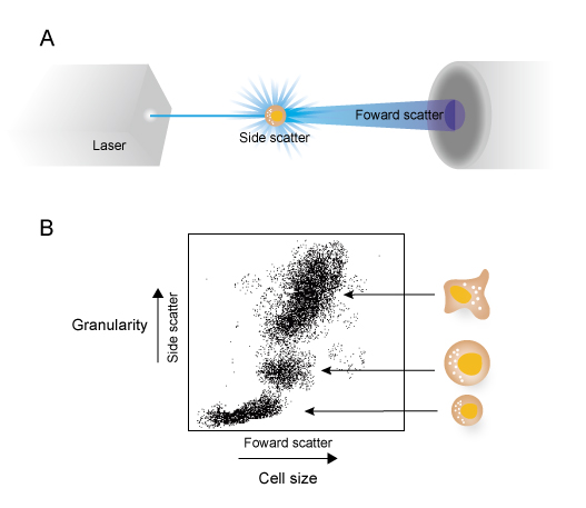
10 Flow Cytometry Principle A Beam Of Light From A Laser Is Passed Download Scientific Diagram

Signal Processing Flow Cytometry Medical Laboratory Science Medical Laboratory

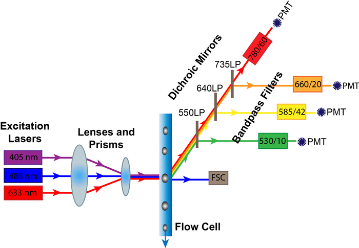
Optical Diagram Of The Facscalibur Benchtop Flow Cytometer From Bd Download Scientific Diagram
Flow Cytometry Guide Creative Diagnostics

Icgeb Facilities
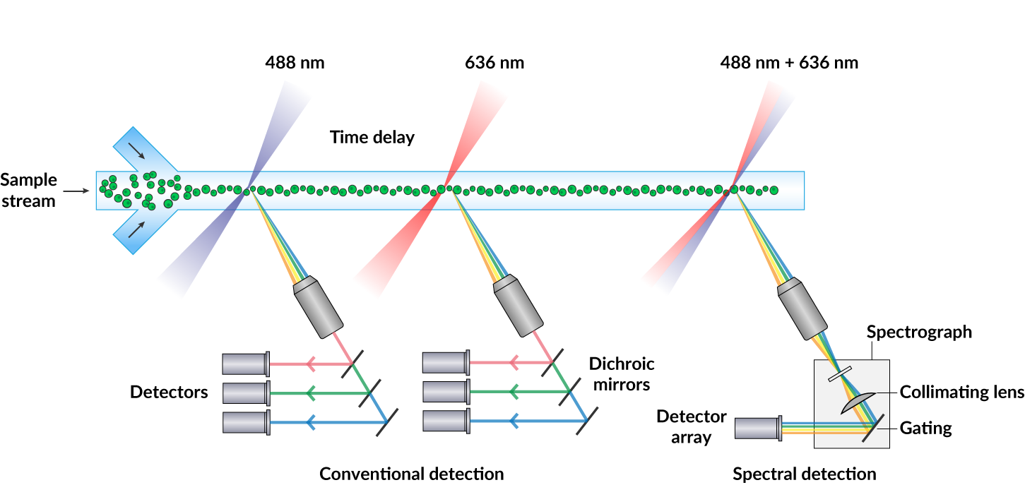
488 nm laser 405 nm laser 633 nm laser 17 ms 55 ms time delay.
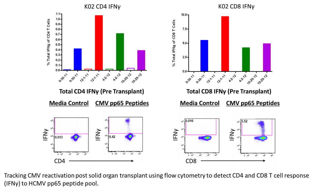
488 nm laser flow cytometry.

Photoacoustic Flow Cytometry Schematics Of The Integrated In Vivo Download Scientific Diagram
Flow Cytometry Fc Protocol Epigentek

Determining Flow Cytometer Resolution Using Multi Peak Capture Beads Flow Cytometry Capture Detection

Reagents For High Parameter Flow Cytometry Bd Biosciences Eu

Spectral Flow Cytometry Fundamentals Thermo Fisher Scientific Ng

Features Optics Fluo
10 Tips For Designing Multi Color Flow Panels Labclinics

Configuration Of The Flow Cytometry Chip The Fluidic Channel Is For 3d Download Scientific Diagram

Flow Cytometery Filters Alluxa
Improving The Flow Cytometry Based Detection Of The Cellular Uptake Of Gold Nanoparticles Analytical Chemistry X Mol

Getting More Research Benefits From Abbkine Innovative Cell Counting Kit 8 Cck 8 Cells Activity Cell Counting

Flow Cytometry Core Facility On The 4th Floor Of The Jimmy Fund Building Flow Cytometry Technology Is Used For Detecting Certain Kinds Of Cells Within Tissue O
Rush Flow Cytometry Core Rush Core Laboratories Rush University

Nkjpwraqwi2clm

Color Wavelengths Chart Google Search Color Wavelengths Color Chart
Plos One Ratiometric Analysis Of Fura Red By Flow Cytometry A Technique For Monitoring Intracellular Calcium Flux In Primary Cell Subsets

Flow Cytometry Panel Design The Basics Thermo Fisher Scientific Ru

How Does It Work Research At St Michael S Hospital
1
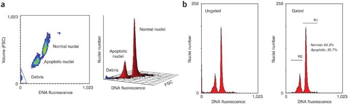
Analysis Of Apoptosis By Propidium Iodide Staining And Flow Cytometry Nature Protocols

Alexa Fluor 488 Anti Cpt1a Antibody 8f6ae9 Ab171449 Abcam

Ultra High Throughput Multiparametric Imaging Flow Cytometry Towards Diffraction Limited Sub Cellular Detection Biorxiv
Spectral Flow Cytometry Biotium

What Is Flow Cytometry Sino Biological
Source : pinterest.com
